
Software from Dassault Systemes offers a 3D image of a patient's heart.


The 3D imaging software that helps Airbus to design planes and Advanced Micro Devices to test processor designs may also aid cardiologists in treating heart disease and reducing health care costs.
Dassault Systemes, long known for its 3D design software used in industries including aerospace, manufacturing and architecture, Tuesday unveiled software that can create 3D models that simulate a human heart. The company is billing the software as the first application with this capability.
"The world flies with our software. The world does not live with our software yet," said CEO Bernard Charlès in an interview.
The health care market isn't entirely new to Dassault Systemes, which is based in Vélizy-Villacoublay, France. Medical equipment and device manufacturers use its software to design and model their products. But this marks Dassault Systemes' entry into using big data to personalize cardiovascular treatments.
The application, which was created using technology found in the company's other 3D software programs, uses a patient's echocardiogram, MRI and CT scan images as well as research data to create a 3D model unique to that person. The technology could help identify potential heart disease risks or allow a surgeon to practice operating before performing a real procedure on a patient.
"If I'm a heart surgeon I don't want to do the operation without having trained myself on your configuration," said Charlès. "How many surgeons are virtual trained? It's still a small percentage. But the virtual training of surgery and complex medical activities will be done using the type of platform that we offer."
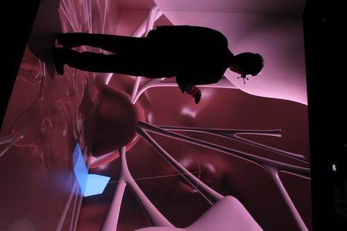
The software was developed as part of the Living Heart Project, which brought together cardiologists, biomechanical specialists and technology professionals to develop 3D modelling technology to help diagnose, prevent and treat heart disease. During a demonstration at Dassault Systemes' U.S. headquarters in Waltham, Massachusetts, the software was run on a tablet-like display from zSpace and in a 3D cave, a three-walled space that also displays images on the floor. In both cases, users wear headsets to experience 3D viewing.
Using a stylus on the tablet, users can zoom in on 3D images of the heart, view individual parts of the heart from different angles, view electrical and mechanical characteristics and view the body's nervous and muscular systems, among other features. The cave offers similar clinical perspectives but presents them in a large, immersive environment. Users can, for example, explore the heart's interior by standing inside its chambers and arteries. A joystick-like device is used to navigate.
If a 3D model is used for a trial run through a surgical procedure, it an also capture detailed explanations on why certain procedures were used during surgery.
"You can't do that with a document because how do you write this down," said Charlès. "It's too difficult to write down. If we make decisions in surgery, two years later it can come back to why we made those decisions. Those experiences are very critical for the future."
Using technology in clinical environments can help control rising health care costs, said Charlès.
The health care sector could learn how to better leverage technology from the aerospace industry, which also produces technically complex products with contributions from a multitude of people.
"Why is it that the plane takes off?" he said. "It's because they are built on a full digital model of everything they do. The health sector is just about discovering that now."
Cardiovascular surgeons realize that using their intuition in conjunction with 3D imaging can produce better and faster results, said Julius Guccione, professor in residence at the University of California, San Francisco School of Medicine, who worked on the Living Heart Project.
"They'll be able to filter out the intuition that really doesn't make sense, doesn't really have a strong physical basis," he said. Given the multiple parameters of open-heart surgery, doctors who are removing heart tissue "really need IT help deciding how much material to remove and from what location," he said. Technology could also help figure out the best time for a procedure and avoid additional operations, he added.
Guccione noted that 3D imaging is already yielding clinical benefits. Another holographic imaging application used in surgery planning reduced the time of a particular operation by more than half, he said.
"That clearly saves costs right there and probably improves outcome if the patient isn't under anesthesia for nearly as long," he said.
Fred O'Connor writes about IT careers and health IT for The IDG News Service. Follow Fred on Twitter at @fredjoconnor. Fred's e-mail address is fred_o'connor@idg.com